


Anti-tumor immune responses can be influenced by a variety of factors ranging from the origin of the cancer cell type, the tissue the tumor arises from, but also from the activation status of the immune system.
Response rates to immunotherapeutic interventions differ dramatically between cancer types ranging from approx. 50% in melanoma to 25% in lung cancer to almost 0% in ovarian cancer. While a lack of response might in part be due to a lack of T cell infiltration, this does not fully explain the differences observed between different cancer types. By analyzing human samples and using genetically-engineered mouse models of ovarian carcinoma, non-small cell lung cancer and skin cancer, we aim to further understand the tumor-derived, tissue macro-environment or systemic environmentally triggered factors that predominantly influence the anti-tumor immunity within the tumor microenvironment.
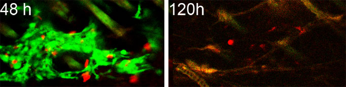
Eradication of tumor lesion mediated by adoptively transferred in vitro-activated, tumor-specific 2C TCR-transgenic T cells. T cells were labeled with dark red fluorescent dye (red) and transferred into mice with melanoma lesions that are driven by BrafV600E and PTEN-deletions (Tyr:CreER, LSLBrafV600E, PTENfl/fl, R26-LSL-SIY, R26-LSL-YFP). Tumor cells (YFP) are illustrated in green. Left, 48 hours post adoptive transfer; right 120h post adoptive transfer. 25x emulsion lens, Leica SP6.
T cells interaction with a T cell-inflamed and a non-T cell-inflamed melanoma
Interaction of adoptively transferred in vitro-activated, tumor-specific T cells (2C TCR-transgenic), labeled with dark red fluorescent dye (red) and transferred into mice with melanoma lesions driven by BrafV600E and PTEN-deletions (Tyr:CreER, LSLBrafV600E, PTENfl/fl, R26-LSL-SIY, R26-LSL-YFP, left) or BrafV600E, PTEN-deletions and activated beta-catenin signaling (Tyr:CreER, LSLBrafV600E, PTENfl/fl, CATLSL-STA, R26-LSL-SIY, R26-LSL-YFP, right). Tumor cells are illustrated in green, 25x emulsion lens, Leica SP6, left 72 hours post adoptive transfer; right 120h post adoptive transfer. In tumors with baseline beta-catenin signaling T cells interact dynamically with tumor cells, a behavior that is not observed in non-T cell-inflamed tumors with activated beta-catenin signaling.
T cells eradicating a T cell-inflamed melanoma
Tumor lesion eradication mediated by adoptively transferred in vitro-activated, tumor-specific T cells (2C TCR-transgenic) that are labeled with dark red fluorescent dye (red) and transferred into mice with melanoma lesions driven by BrafV600E and PTEN-deletions (Tyr:CreER, LSLBrafV600E, PTENfl/fl, R26-LSL-SIY, R26-LSL-YFP). Tumor cells are illustrated in green, 25x emulsion lens, Leica SP6, left 48 hours post adoptive transfer; right 120h post adoptive transfer.
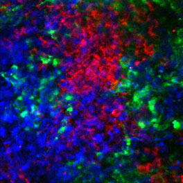
Tumor (B16) cells expressing green fluorescent protein or red fluorescent protein injected into a host expressing cerulean fluorescent protein in immune and stroma cells. This nicely illustrates the intimate interaction between tumor cells and other cells of the tumor-immune microenvironment. Image taken 6 days post tumor injection, 25x emulsion lens, Leica SP6.